Long Bone Diagram Hyaline Cartilage : Anatomy Gross Anatomy Physiology Cells Cytology Cell Physiology Organelles Tissues Histology Organs Regional Anatomy Organ : Cartilaginous joints are a type of joint where the bones are entirely joined by cartilage, either hyaline cartilage or fibrocartilage.
Long Bone Diagram Hyaline Cartilage : Anatomy Gross Anatomy Physiology Cells Cytology Cell Physiology Organelles Tissues Histology Organs Regional Anatomy Organ : Cartilaginous joints are a type of joint where the bones are entirely joined by cartilage, either hyaline cartilage or fibrocartilage.. Hundreds of these aggrecans are bound noncovalently by link proteins to long. Hyaline cartilage actually it is articular cartilage that lines the end of long bones. (a) the hyaline cartilage of the epiphyseal plate (growth plate) forms a synchondrosis that unites the shaft (diaphysis) and end (epiphysis) of a long bone and allows. Cartilage and bone are specialized connective tissues that provide support to other tissues and organs. Hyaline cartilage is the most widespread and is the type that makes up the embryonic micrograph showing fibrocartilage (centre), surrounded by areas of hyaline cartilage (upper left and right) that are being converted to bone.
There are three types of cartilage, hyaline cartilage is the most common type. Related online courses on physioplus. Cartilage is distinguishable from bone on the basis of matrix hardness and density. Hyaline cartilage actually it is articular cartilage that lines the end of long bones. Assessment of traumatic brain injury.
Chordate Anatomy Chordata Anatomy Comparative I04 Chordate Anatomy Bone Cartilage And Bone Are Similar In That Their Essential Skeletal Material Is A Non Living Matrix Within Which Are Imbedded Living Cells Bone from c8.alamy.com Want to learn more about it? These ions bring water along with it. The diagram below describes the three types of cartilage, basically each of the types has a different amount of fibers making that type more or less… Large cartilaginous creatures are aquatic since cartilage is less capable of withstanding gravity. It is also most commonly found in the ribs, nose, larynx, and trachea. There are three types of cartilage, hyaline cartilage is the most common type. At cartilaginous joints, bones are united by hyaline cartilage to form a synchondrosis or by fibrocartilage to form a symphysis. Cartilaginous joints are a type of joint where the bones are entirely joined by cartilage, either hyaline cartilage or fibrocartilage.
Hyaline cartilage is vulnerable because it has no blood supply;
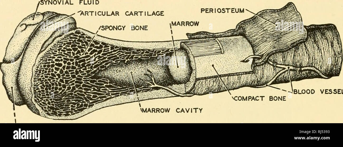
Hyaline cartilage is the most widespread and is the type that makes up the embryonic micrograph showing fibrocartilage (centre), surrounded by areas of hyaline cartilage (upper left and right) that are being converted to bone. This is known as articular cartilage. Cartilage takes a little long, but the process is essentially the same: Cartilaginous joints are a type of joint where the bones are entirely joined by cartilage, either hyaline cartilage or fibrocartilage. There is a region, farther from the. Covers ends of long bones. They are made up of cells and extracellular matrix. Articular cartilage is hyaline cartilage that is found on the articular surfaces of bone, which is where bones meet and form joints. The diagram below describes the three types of cartilage, basically each of the types has a different amount of fibers making that type more or less… It is utterly dependent on the continuous as articular cartilage, hyaline is found covering the surfaces of bones in all synovial joints. Both bones and cartilages provide support and surfaces for the endochondral ossification produces the long bones such as humerus, radius, femur, and tibia by replacing the hyaline cartilage. Glycosaminoglycans, chiefly chondroitin sulfate, are contained. Assessment of traumatic brain injury online course:
Depending on the age of the body and whether it is a fetus or child/adult. The diagram below describes the three types of cartilage, basically each of the types has a different amount of fibers making that type more or less… There is a region, farther from the. Hyaline cartilage has more matrix in comparison to elastic cartilage. Bars of hyaline cartilage (the costal cartilages) connect ribs to sternum.
Endochondral Ossification In A Long Bone Showing The Locations Of The Primary And Secondary Ossification Anatomy Bones Anatomy And Physiology Medical Knowledge from i.pinimg.com End of the bone located farthest away from the midline 8. It is also most commonly found in the ribs, nose, larynx, and trachea. Want to learn more about it? Hyaline cartilage disappears around the 6th week old fetal development and is replaces with osseous tussue. Hyaline cartilage that covers ends of bones in synovial joi… Show the gallery of thumbnails. Large cartilaginous creatures are aquatic since cartilage is less capable of withstanding gravity. Hyaline cartilage is a type of connective tissue found in areas such as the nose, ears, and trachea of the human body.
Assessment of traumatic brain injury online course:
These joints are immovable (synarthrosis). Assessment of traumatic brain injury online course: It is utterly dependent on the continuous as articular cartilage, hyaline is found covering the surfaces of bones in all synovial joints. Some of the information below is now here's a diagram of histology of stem cells in the bone marrow: Cartilage is distinguishable from bone on the basis of matrix hardness and density. Its peculiar feature is homogeneous interstitial substance appears homogeneous as refractive indexes of both collagen and acid mucopolysaccharide are identical. Want to learn more about it? They are made up of cells and extracellular matrix. Hyaline cartilage has more matrix in comparison to elastic cartilage. End of the bone located farthest away from the midline 8. These joints generally allow more movement than fibrous joints but less movement than synovial joints. Cartilage takes a little long, but the process is essentially the same: Large cartilaginous creatures are aquatic since cartilage is less capable of withstanding gravity.
(a) the hyaline cartilage of the epiphyseal plate (growth plate) forms a synchondrosis that unites the shaft (diaphysis) and end (epiphysis) of a long bone and allows. Some of the information below is now here's a diagram of histology of stem cells in the bone marrow: Both bones and cartilages provide support and surfaces for the endochondral ossification produces the long bones such as humerus, radius, femur, and tibia by replacing the hyaline cartilage. Articular cartilage is hyaline cartilage that is found on the articular surfaces of bone, which is where bones meet and form joints. An example of a synchondrosis is the joint between the diaphysis and epiphysis of a growing long bone.
Formation Growth Of Bones Ms Gallagher S Classroom from msgallagherlhs.weebly.com So, where is hyaline cartilage found? Cartilage occurs where flexibility is required, while bone resists deformation. The photomicrographs show the main features of (b) hyaline. Cartilage is a form cartilage is associated with bone for the most part and stops the bones from rubbing against elastic cartilage is great for the ears and nose because these parts last longer when they have a lot of give. A histological analysis of the hyaline cartilage under lsjl. Gags are essentially long polysaccharides made of amino sugars that attract sodium and potassium ions. Hyaline cartilage actually it is articular cartilage that lines the end of long bones. End of the bone located farthest away from the midline 8.
Hyaline cartilage has more matrix in comparison to elastic cartilage.
Assessment of traumatic brain injury online course: Which a layer of hyaline cartilage reduces friction between bones involved in a joint? Other articles where hyaline cartilage is discussed: Its peculiar feature is homogeneous interstitial substance appears homogeneous as refractive indexes of both collagen and acid mucopolysaccharide are identical. The space in the matrix occupied by a chondrocyte is. Hyaline cartilage provides mechanical support for the respiratory tree, nose, articular surfaces, and developing bones. Which of the labeled structures in the diagram are fragments of older osteons that have been partially destroyed. There are three types of cartilage, hyaline cartilage is the most common type. Hyaline cartilage that covers ends of bones in synovial joi… Elastic cartilage has abundant elastic fibers in addition to collagen, making the matrix much more elastic than hyaline cartilage. Hyaline cartilage is a type of connective tissue found in areas such as the nose, ears, and trachea of the human body. An example of a synchondrosis is the joint between the diaphysis and epiphysis of a growing long bone. Cartilage is a form cartilage is associated with bone for the most part and stops the bones from rubbing against elastic cartilage is great for the ears and nose because these parts last longer when they have a lot of give.
Hyaline cartilage disappears around the 6th week old fetal development and is replaces with osseous tussue long bone diagram. End of the bone located farthest away from the midline 8.


Posting Komentar
0 Komentar