Blood Vessels Labeled Brain - Blood Vessels of the Brain & Spinal Cord - Atlas of Anatomy : Capillaries are blood vessels that are one cell thick (endothelium) where the main diffusion and exchange takes place.
Blood Vessels Labeled Brain - Blood Vessels of the Brain & Spinal Cord - Atlas of Anatomy : Capillaries are blood vessels that are one cell thick (endothelium) where the main diffusion and exchange takes place.. The wire core is made from nitinol, a nickel titanium alloy. He says the restricted vessels prevent the blood from draining fast enough and injure the brain by causing a build up of iron which leads to ms. Blood travels from the heart in arteries, which branch into smaller and smaller vessels, eventually becoming arterioles. Blood vessels in red in close communication with proliferating neuronal cells in the mouse cortex at embryonic day 10. Blood vessels flow blood throughout the body.
Cells that line the walls of blood vessels). Blood vessels in red in close communication with proliferating neuronal cells in the mouse cortex at embryonic day 10. This vessel supplies blood to the front part of your brain, knows as your frontal lobe. The endothelium is known as the tunica intima. Where venules are smaller versions of veins.
Vertebral Artery Segments, Stenosis and Artery Dissection ... from healthjade.com A blood clot that lodges in a brain blood vessel, causing a stroke. However, they have observed blood vessel damage caused. Sudden interruption of blood flow and oxygen to an area of brain tissue, which then may die (cerebrovascular accident, or cva, is another name for stroke.) ischemic stroke: The capillaries also connect the branches of arteries and to. Posterior communicating a internal carotid а. The carotid arteries and the vertebral arteries anterior cerebral artery (aca): Identify all of the blood vessels that are illustrated in the figure as you can while holding or otherwise examining whole brain specimens. Examine a second specimen and notice any differences, such as asymmetries in the size of the vertebral or posterior communicating arteries.
A blood clot that lodges in a brain blood vessel, causing a stroke.
There is a right sided aca and a left sided aca. Veins are blood vessels that return blood back to the heart; In this video i discuss the major arteries that supply the brain, starting with the internal carotid and vertebral arteries and covering many of the major. The wire core is made from nitinol, a nickel titanium alloy. Blood travels from the heart in arteries, which branch into smaller and smaller vessels, eventually becoming arterioles. He says the restricted vessels prevent the blood from draining fast enough and injure the brain by causing a build up of iron which leads to ms. These vessels transport blood cells, nutrients, and oxygen to the tissues of the body. Blood vessels are intricate networks of hollow tubes that transport blood throughout the entire body so that it can deliver valuable nutrients to and remove waste from cells. Identify all of the blood vessels that are illustrated in the figure as you can while holding or otherwise examining whole brain specimens. Supplies the posterior brain, blood supply to the entire brain is ensured by anastomoses between the vessels. Posterior communicating a internal carotid а. Traditionally, pais has been explained as being caused by a blood clot forming within the ageing placenta, entering the fetal circulation, embolising across the patent foramen ovale, travelling into the left ventricle, into the ascending aorta and then one of the main three branches of the thoracic aorta. Blood is also supplied to the brain by the vertebral a.
Supplies the anterior brain and the vertebral a. The capillaries also connect the branches of arteries and to. Blood vessels flow blood throughout the body. Traditionally, pais has been explained as being caused by a blood clot forming within the ageing placenta, entering the fetal circulation, embolising across the patent foramen ovale, travelling into the left ventricle, into the ascending aorta and then one of the main three branches of the thoracic aorta. Sudden interruption of blood flow and oxygen to an area of brain tissue, which then may die (cerebrovascular accident, or cva, is another name for stroke.) ischemic stroke:
Skull Arteries | Bruin Blog from o.quizlet.com The dense tight junctions between endothelial cells prevent paracellular transport through the. Choose from 500 different sets of flashcards about mastering a&p anatomy of blood vessels label veins on quizlet. Scientists are developing new strategies for attaching drugs to molecules naturally transported across the barrier (labeled in green and blue). A blood clot that lodges in a brain blood vessel, causing a stroke. Veins are blood vessels that return blood back to the heart; The endothelium is known as the tunica intima. Posterior communicating a internal carotid а. Blood vessels are referred to collectively as the vascular system and, together with the heart, make up the circulatory system or cardiovascular system.
Another whole article within the blood vessels and csf section is dedicated to the cavernous sinus.
The dense tight junctions between endothelial cells prevent paracellular transport through the. Traditionally, pais has been explained as being caused by a blood clot forming within the ageing placenta, entering the fetal circulation, embolising across the patent foramen ovale, travelling into the left ventricle, into the ascending aorta and then one of the main three branches of the thoracic aorta. The endothelium is known as the tunica intima. Uc davis institute for regenerative cures. These vessels transport blood cells, nutrients, and oxygen to the tissues of the body. The carotid arteries and the vertebral arteries anterior cerebral artery (aca): Internal carotid artery (anterior circulation), vertebral artery (posterior circulation), and their hexagonal anastomotic network called blood brain barrier refers to the wall between the brain tissue and blood vessels. Only some of the vessels that exist in a real brain have been labeled. Cerebral arterial circle anterior communicating posterior cerebral a middle cerebral al reset zoom. Arteries transport blood away from the heart. Hydrogels are formed from biocompatible polymers. Blood vessel endothelium is continuous with the inner tissue lining of organs such as the brain, lungs, skin, and heart. The difference in the structural characteristics of arteries, capillaries and veins is attributable to their respective functions.
Blood vessels in red in close communication with proliferating neuronal cells in the mouse cortex at embryonic day 10. Cerebral arterial circle anterior communicating posterior cerebral a middle cerebral al reset zoom. Choose from 500 different sets of flashcards about mastering a&p anatomy of blood vessels label veins on quizlet. These structures are responsible for the production, transport and removal of. However, they have observed blood vessel damage caused.
Corrosion cast of brain blood vessels, pl | Wellcome ... from i.pinimg.com The 500 ms patients, both adults and children, also underwent mri scans of the brain to measure iron deposits in surrounding areas of the brain. Blood travels from the heart in arteries, which branch into smaller and smaller vessels, eventually becoming arterioles. Supplies the posterior brain, blood supply to the entire brain is ensured by anastomoses between the vessels. The wire core is made from nitinol, a nickel titanium alloy. This is particularly important structure due to its the ventricular system is a set of communicating cavities within the brain. • identification of blood vessels as arteries, capillaries or veins from the structure of their walls. Blood vessels in red in close communication with proliferating neuronal cells in the mouse cortex at embryonic day 10. Capillaries are blood vessels that are one cell thick (endothelium) where the main diffusion and exchange takes place.
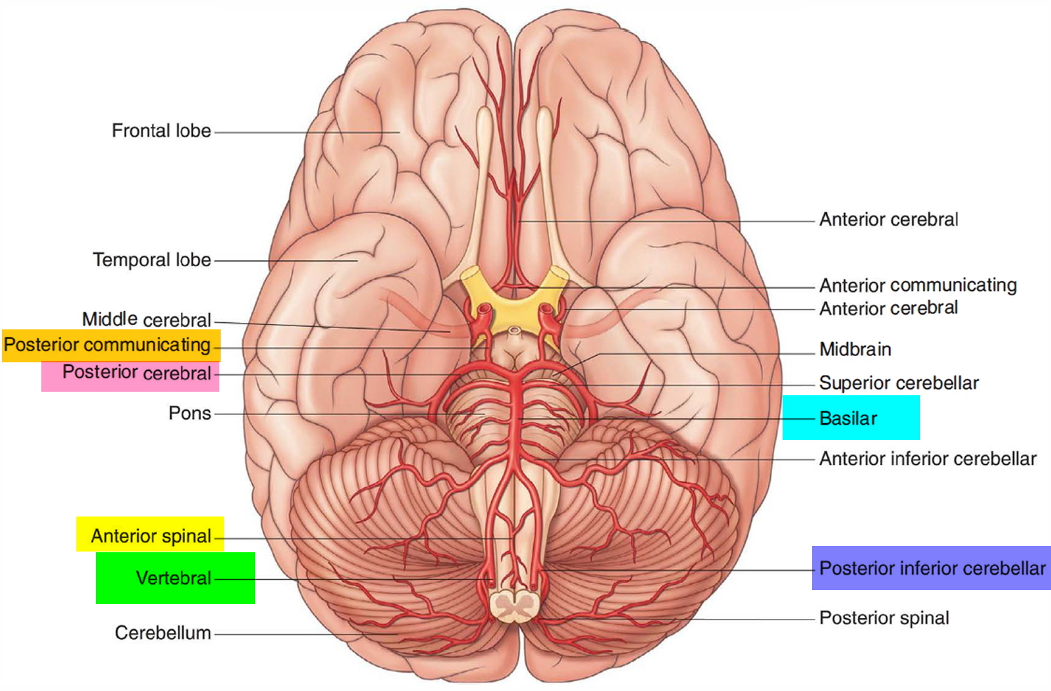
Blood in the brain is supplied by two pairs of large blood vessels (arteries):
This is particularly important structure due to its the ventricular system is a set of communicating cavities within the brain. Blood vessels are vital for the body and play a key role in diabetes helping to transport glucose and insulin. The blood vessels (and nerves) enter the brain through holes in the skull called foramina. The blood vessels are the components of the circulatory system that transport blood throughout the human body. Label the blood vessels in the inferior view of the brain using the hints provided. Supplies the posterior brain, blood supply to the entire brain is ensured by anastomoses between the vessels. The capillaries also connect the branches of arteries and to. Internal carotid artery (anterior circulation), vertebral artery (posterior circulation), and their hexagonal anastomotic network called blood brain barrier refers to the wall between the brain tissue and blood vessels. Capillaries are blood vessels that are one cell thick (endothelium) where the main diffusion and exchange takes place. There is a right sided aca and a left sided aca. Another whole article within the blood vessels and csf section is dedicated to the cavernous sinus. They also take waste and carbon dioxide away from the tissues. Blood vessels in red in close communication with proliferating neuronal cells in the mouse cortex at embryonic day 10.
Growing brains in laboratories was just the start for scientists blood vessels labeled. Blood vessels are referred to collectively as the vascular system and, together with the heart, make up the circulatory system or cardiovascular system.


Posting Komentar
0 Komentar